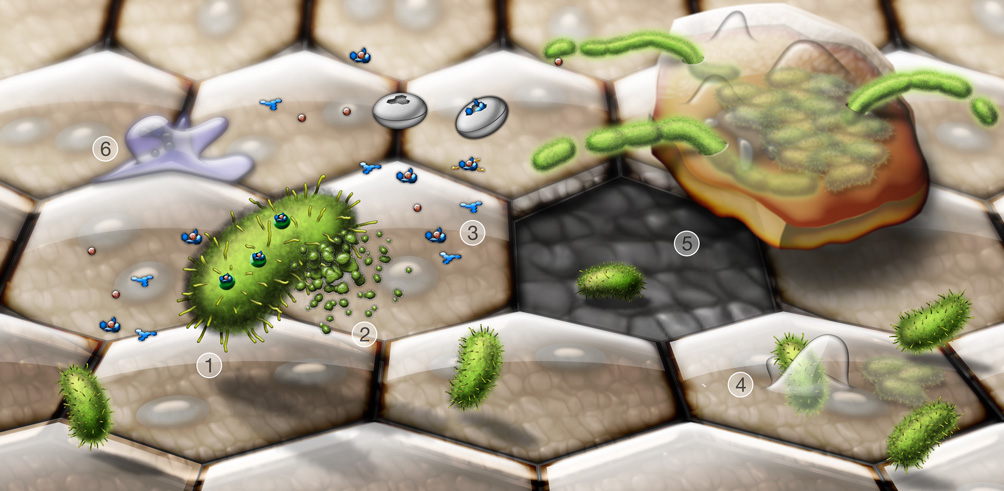
Figure 1.
Dynamic interplay between invading UPEC and the host during a UTI. Shown are key events taking place during bladder infection by UPEC. Type 1 pili-expressing UPEC (1, green) secrete toxins and other virulence factors, alone or in association with outer membrane vesicles (2). Siderophores like enterobactin and salmochelin (3, blue structures) released by UPEC scavenge iron, in competition with host iron chelating molecules and lipocalin 2 (white discs). Type 1 pili mediate bacterial attachment to and invasion of the bladder epithelial cells (4). Large terminally differentiated superficial epithelial cells, which are often binucleate and have distinctive hexagonal or pentagonal shapes, line the lumenal surface of the bladder and are the primary targets of UPEC invasion. UPEC can rapidly multiply within the superficial cells, forming large biofilm-like communities. Exfoliation of infected bladder cells facilitates bacterial clearance from the host, but leaves the smaller underlying immature cells more susceptible to infection (5). The release, or efflux, of UPEC from infected host cells before they complete exfoliation likely promotes bacterial dissemination and persistence within the urinary tract. During efflux, UPEC often become filamentous, probably due in part to mounting stress arising from increased activation of host defenses. These include the influx of neutrophils (6), as well as the generation of reactive oxygen and nitrogen species and antimicrobial peptides.

