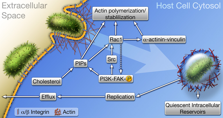
Figure 3.
Host cell invasion by UPEC. The FimH adhesin localized at the distal tips of type 1 pili engages α3β1 integrin receptors, and possibly other receptors, which likely cluster within cholesterol-rich lipid rafts. Receptor binding triggers signaling cascades involving FAK, Src, PI 3-kinase, Rho GTPases like Rac1, phosphoinositides (PIPs), and transient complex formation between the cytoskeleton stabilizing and scaffolding proteins α-actinin and vinculin. These events stimulate actin rearrangements, causing the host plasma membrane to zipper around and envelope bound bacteria. Once internalized, UPEC can be trafficked to late endosome-like compartments that are often localized within a meshwork of actin filaments. Bacteria exist quiescently within these actin-bound compartments and may serve as reservoirs for recurrent UTIs. Liberation of UPEC into the host cytosol stimulates rapid bacterial growth and the formation of intracellular biofilm-like communities.

