Figure 2.

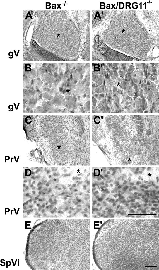
Photomicrographs of horizontal sections through the gV (A–B′) and coronal sections through the PrV (C–D′) and SpVi (E, E′) from Bax−/− and Bax/DRG11 double −/− cases on the day of birth. All conventions are as in Figure 1.

Photomicrographs of horizontal sections through the gV (A–B′) and coronal sections through the PrV (C–D′) and SpVi (E, E′) from Bax−/− and Bax/DRG11 double −/− cases on the day of birth. All conventions are as in Figure 1.