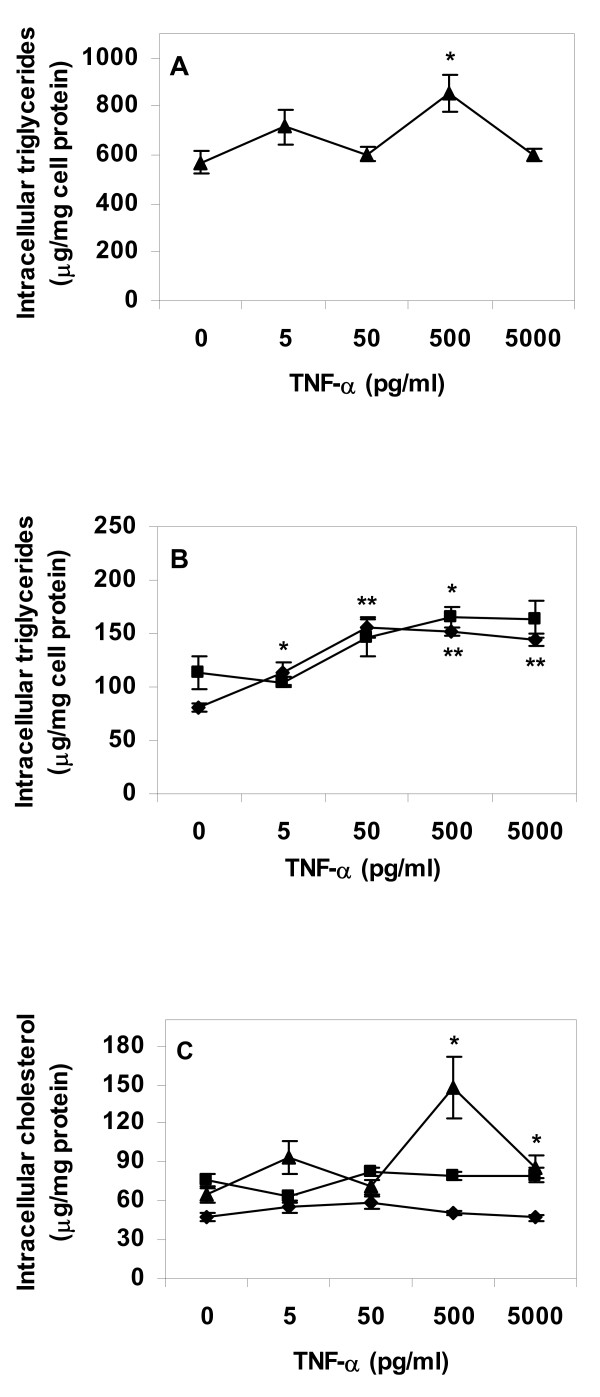
Figure 2.
Lipid content of primary human macrophage incubated with TNF-α. Cells were differentiated for four days with GM-CSF, and then incubated for 24 h in absence or presence of lipoproteins (50 μg/ml), rinsed with heparin and incubated with TNF-α in lipoprotein-free media for an additional 24 h. Intracellular lipids were extracted and lipid values normalized to cell protein content. A. Triglyceride content of VLDL treated cells, B. Triglyceride content of control or AgLDL treated cells, C. Cholesterol content of cells. Data represents mean ± SEM (n = 6) for a representative experiment repeated three times with cells from different donors. * = P < 0.05, ** = P < 0.01 compared to cells incubated in absence of IL-1β. Diamonds = control cells (no lipoprotein lipid loading), squares = AgLDL, triangles = VLDL.

