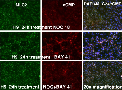
Fig. 5.
Immunofluorescence detection of MLC2 and cGMP in H-9 cells exposed to NO donor and sGC activator. Differentiated cells were exposed to NOC-18 (1 μM; Top), BAY 41-2272 (3 μM; Middle), or the combination of the two (C) for 24 h at 37 °C. The cells were fixed with paraformaldehyde and incubated with antibodies to MLC2 and cGMP that was followed by detection with fluorescent-conjugated secondary anti-mouse or rabbit antibodies. (Magnification: 20×.)

