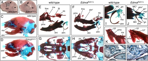
Fig. 1.
Transformation of maxillary components in EdnraEdn1/+ mice. (A and B) Facial appearance of E18.5 wild-type (A) and EdnraEdn1/+ (B) mice. Vibrissae are absent in mutant mice that have open eyelids (arrows) and an anterior shift of the ear position (arrowheads). (C–L) Bone and cartilage elements of E18.5 wild-type (C, E, G, I, and K) and EdnraEdn1/+ (D, F, H, J, and L) skulls. (C and D) Lateral views of wild-type and EdnraEdn1/+ craniofacial skeletons. (E and F) The EdnraEdn1/+ mutant shows mirror-image duplication of the mandibular elements (dentary, MC, and malleus) at the expense of the maxillary elements (maxilla, jugal, squamosal, and incus). (G and H) Caudal views of wild-type and EdnraEdn1/+ craniofacial skeletons. The dentaries are removed to show the normal and transformed maxillae. Large parts of the palatine, pterygoid, lamina obturans, and ala temporalis are missing or severely deformed in the EdnraEdn1/+ mutant. (I and J) Ectotympanic and gonial bones are also duplicated in association with the ectopic MC in EdnraEdn1/+ mutants. (K and L) Caudal views of the premaxilla-maxilla junction. The tip of the transformed maxilla-dentary in the EdnraEdn1/+ mutant contains an ectopic incisor and fuses to the premaxilla. (M and N) Parasagittal sections of E18.5 wild-type (M) and EdnraEdn1/+ (N) mice stained by Mallory trichromic. The EdnraEdn1/+ mutant shows an ectopic incisor in addition to the orphotopic 1. Fusion between the premaxilla and transformed maxilla-dentary is observed in the mutant. at, ala temporalis; bs, basisphenoid; dnt, dentary; etm, ectotympanic; fmx, frontal process of maxilla; gn, gonial; hy, hyoid; in, incus; jg, jugal; lo, lamina obturans; ma, malleus; mx, maxilla; pl, palatine; pmx, premaxilla; ps, presphenoid; ptg, pterygoid; sq, squamosal; tg, tongue; UI, upper incisor; vbf, vibrissae follicle; vm, vomer; *, ectopic structure.

