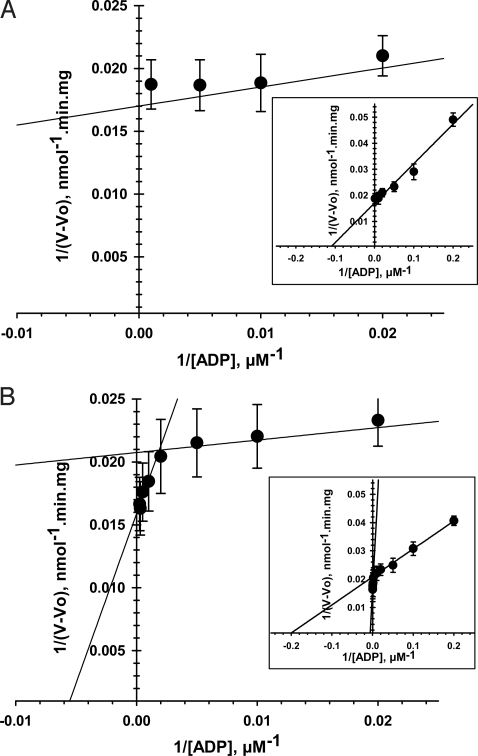
Fig. 5.
Tubulin dramatically increases apparent Km for ADP in regulation of respiration of isolated brain mitochondria. Shown are double-reciprocal representations of the respiration kinetics of brain mitochondria activated by ADP in control (A) and in the presence of 1 μM tubulin (B). The 2 straight lines represent 2 different respiration kinetics in the presence of tubulin (B). (Insets) Enlargements of A and B. Each data point is a mean of 6–9 independent experiments ±SE.

