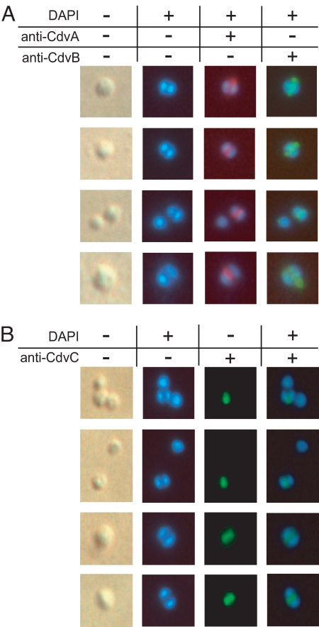
Fig. 2.
In situ immunofluorescence microscopy of S. acidocaldarius cells. Cultures were sampled in exponential growth phase. The first column depicts phase-contrast illumination of the cells shown in the consecutive columns. Nucleoids were stained with DAPI (4′,6-diamidino-2-phenylindole). Cdv proteins were stained with specific antibodies, followed by fluorescence visualization with Alexa Fluor-labeled secondary antibodies, as described in Materials and Methods. (A) Cells with two segregated nucleoids double-stained with anti-CdvA (red fluorescence) and anti-CdvB (green). (B) Anti-CdvC (green) stained cells. Note the absence of fluorescence signals in single-nucleoid cells (top two rows).

