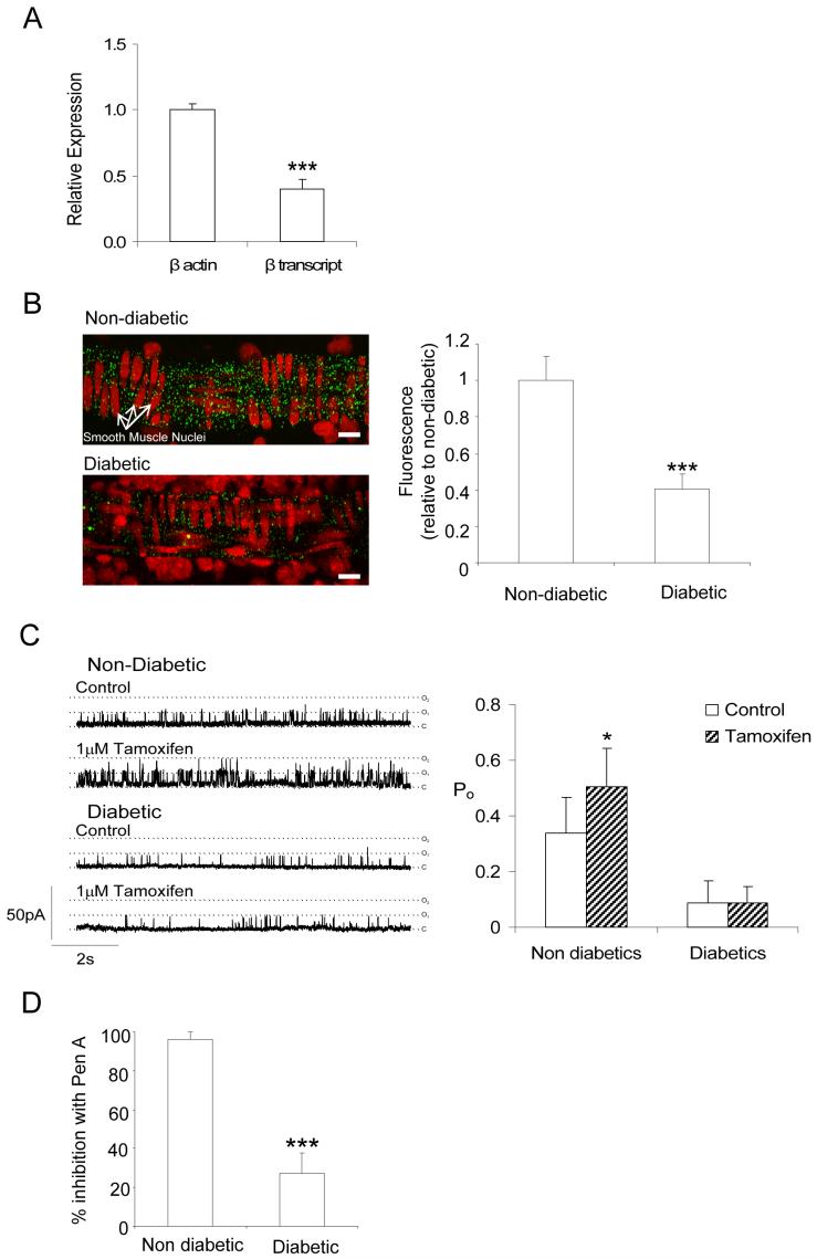
Figure 7. BKβ1 subunit expression and function.
(A) Downregulation of BKβ1 mRNA in retinal VSMC cells from diabetic arterioles. BKβ1 expression in diabetic arterioles is presented relative to non-diabetic vessels. Amplifications were performed in triplicate (same samples as for Fig 5A) and normalized as described for BKα transcripts (B) Left, confocal images of non-diabetic and diabetic retinal arterioles embedded within retinal flatmount preparations and labeled with anti-BKβ1 Ab (green) and propidium iodide (red: nuclear label). Labeling of the circular smooth muscle is reduced in the tissue from the diabetic animal. Right, summary data showing statistically significant reduction in anti-BKβ1 fluorescence for diabetic samples (n=6 retinas, 30 vessels) relative to non-diabetics (n=6 retinas, 25 vessels). (C) Sensitivity of single BK channels in inside out patches to 1μmol/L tamoxifen (holding potential +80mV; 1μmol/L free [Ca2+]) from non-diabetic and diabetic retinal VSMCs. Right, summary data showing the differential effects of tamoxifen on the Po of single BK channels from non-diabetic (n=7) and diabetic (n=8) vessels. (D) Pharmacology of single BK channels from non-diabetic (n=5) and diabetic (n=9) retinal VSMCs exposed to Pen A. Mean data is expressed as the % inhibition of Po after 5-min of exposure to 100nmol/L Pen A.

