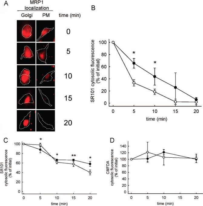
Figure 3.
Comparative assessment of the function of Golgi apparatus versus PM-localized MRP1-EGFP. (A) Visual comparisons of SR101 distribution in HeLa cells as a function of time that have MRP1-EGFP localized at the Golgi apparatus or at the PM. Cell outlines are shown with dashed lines, and the scale bar represents 10 μm. (B) SR101 cytosolic fluorescence as a function of time in HeLa cells transfected with MRP1-EGFP. Cells treated with monensin (●) had MRP1 localized at the Golgi apparatus. Untreated cells (○) had MRP1 at the PM. Values are shown as mean ± SD (n = 9 independent evaluations). Statistical analysis was performed using an unpaired t test (*, p < 0.05). (C) The influence of monensin treatment on SR101 cytosolic fluorescence as a function of time in Hela cells that do not contain MRP1. Cells treated with monensin (●) or left untreated (○) are shown. Values are shown as mean ± SD (n = 9 independent evaluations). Statistical analysis was performed using an unpaired t test (*, p < 0.05; **, p < 0.01). (D) Cytosolic fluorescence of CMFDA-treated cells as a function of time in the absence (●) and presence (○) of monensin. Values are shown as mean ± SD (n = 9 independent evaluations). No statistically significant differences were observed between the groups.

