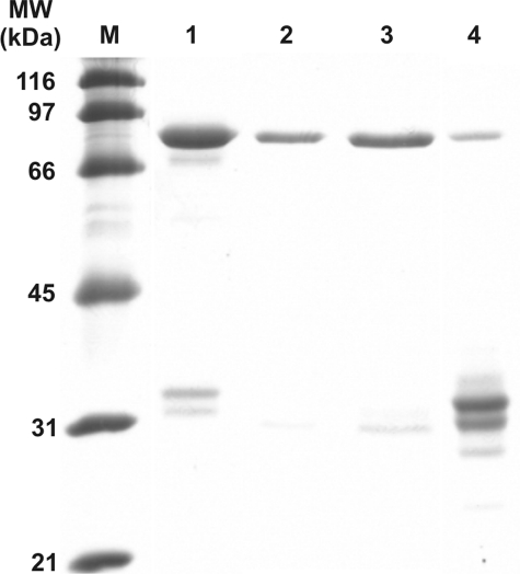
FIGURE 5.
SDS-PAGE analysis of purified recombinant GST human CBS fusion proteins. 10 μg of each protein was separated on 9% SDS-PAGE gel and stained with SimplyBlue SafeStain (Invitrogen). Lanes: M, broad range SDS-PAGE marker (Bio-Rad); 1, Fe(III)-PPIX hCBS; 2, Mn(III)-PPIX hCBS; 3, Co(III)-PPIX hCBS; 4, GST-hemeless hCBS fusion proteins. The small bands visible in lane 4 when examined on a Western blot are not detectable with anti-CBS or anti-GST antibody.

