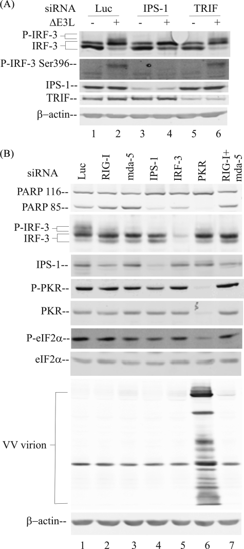
FIGURE 5.
PKR-mediated IRF-3 phosphorylation occurs within the RIG-like receptor-IPS adapter signal transduction pathway. A, whole cell extracts were prepared from parental PKR+ cells, either uninfected (–) or ΔE3L virus-infected (+) for 10 h following transient knockdown as described under “Experimental Procedures,” utilizing chemically synthesized siRNAs against luciferase as a control (lanes 1 and 2), IPS-1 (lanes 3 and 4), or TRIF (lanes 5 and 6). Western immunoblot analyses were carried out with antibodies against IRF-3, phospho-IRF-3(Ser-396), TRIF, IPS-1, and β-actin as a loading control. B, whole cell extracts were prepared from parental PKR+ cells infected with ΔE3L mutant virus following transient knockdown utilizing siRNAs against the following targets: lane 1, luciferase control; lane 2, RIG-I; lane 3, mda-5; lane 4, IPS-1; lane 5, IRF-3; lane 6, PKR; lane 7, RIG-I and mda-5. Western immunoblot analyses were carried out with extracts prepared at 10 h after infection with antibodies against IRF-3, IPS-1, phospho-PKR (P-PKR), PKR, phospho-eIF-2α (P-eIF-2α), and eIF-2α or 24 h after infection to measure PARP cleavage (PARP 85), vaccinia virus protein expression, and β-actin.

