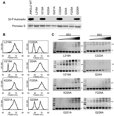
FIGURE 4.
Alanine Scanning Mutagenesis of the JNK2α2 α-region. A, in vitro kinase assay using radioactive [32P]ATP with JNK2α2 wild type and the alanine mutants. The indicated amino acid in JNK2α2 was mutated to alanine. 1 μg of purified fusion protein was then used in an in vitro kinase/autophosphorylation assay. Top panel, autoradiogram of autophosphorylation reactions. In vitro kinase reactions were electrophoresed in SDS-PAGE and the subsequent Western blot used for autoradiography. Bottom panel, Ponceau S stain of the same Western blot to demonstrate equal loading of the respective JNK mutants. B, gel filtration of alanine mutants. Purified fusion proteins for the same mutants were used in size exclusion chromatography. 1-ml fractions were collected, and the A280 nm value was measured. C, cross-linking analysis using increasing concentrations of the homo-bifunctional cross-linker BS3. * indicates the JNK2α2 monomer. ** indicates the JNK2α2 dimer.

