FIGURE 6.
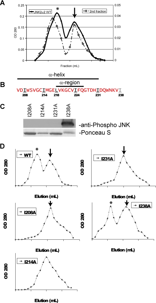
JNK2α2 dimerization may occur through a leucine zipper motif. A, gel filtration of wild type JNK2α2. The left axis represents the initial filtration of WT JNK2α2 in which 1-ml fractions were collected, and the A280 nm was measured. *, JNK2α2 dimer; arrow, monomer. The right axis is the subsequent gel filtration of the dimer fraction. B, amino acid sequence of JNK2α2 indicating the location of the α-region and α-helix. Red letters indicate amino acids that were not mutated. Black/underlined letters indicate amino acids that were mutated to an alanine and resulted in loss of JNK2α2 autophosphorylation. The gray/underlined letter indicates an amino acid that was mutated to an alanine but retained autophosphorylation capability. C, in vitro kinase reactions with the mutant His-JNK2α2. Western blots were probed with an antibody specific for phosphorylated JNK at the T-P-Y motif. Ponceau S stain of the same Western blot demonstrated equal loading of the respective JNK mutants. D, gel filtration of JNK2α2 wild type and JNK2α2 mutants. 1-ml fractions were collected, and the A280 nm values were measured. *, JNK2α2 dimer; arrow, monomer.

