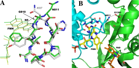
FIGURE 2.
A, superposition of the FMN binding loops in rat nNOS (green) and in cytochrome P450BM3 (white). The hydrogen bonds from the N5 position of FMN to the nearby protein backbone are depicted by dashed lines. The loop in nNOS (green) is a double β-turn, whereas the shorter one in P450BM3 has only a single β-turn. B, the interface between the FMN and FAD binding domains (in cyan and green, respectively) of the nNOS reductase domain (1F20). The hydrogen bond from Asn-811 to Glu-1392 is one of the inter-domain interactions. The H-bond would be disrupted when Gly-810 is deleted in the ΔG810 mutant. The figure was made with PyMOL (W. L. DeLano (2002) PyMOL, DeLano Scientific, San Carlos, CA).

