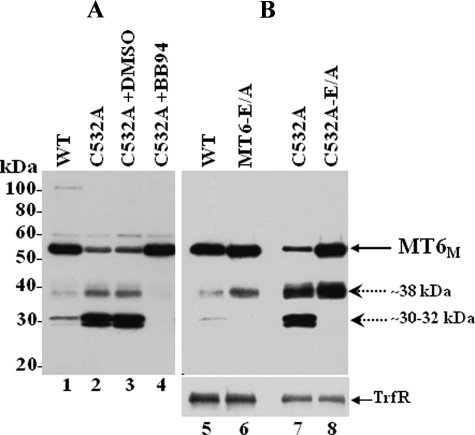
FIGURE 7.
Degradation of MT6-MMP monomers. A, COS-1 cells were transiently transfected to express wild type (WT; lane 1) or C532A (lanes 2–4) MT6-MMP. Six hours after transfection, the cells expressing C532A (lanes 2–4) were incubated without (lane 2) or with 10 μm BB-94 (lane 4) or vehicle (DMSO, lane 3) in complete media. Forty hours post-transfection the cells were harvested, and the lysates were resolved by reducing 10% SDS-PAGE followed by immunoblot analyses with MT6-MMP antibodies. B, COS-1 cells were transiently transfected to express wild type (lane 5), MT6-E/A (lane 6), C532A (lane 7), or C532A-E/A (lane 8) MT6-MMP. Forty-hours later the cells were surface-biotinylated as described under “Experimental Procedures.” The cells were then lysed in cold lysis buffer, and protein concentrations were determined. Equal amounts of protein were subjected to Neutravidin beads pulldown assays. The bound biotinylated proteins were eluted with Laemmli SDS-sample buffer containing β-ME and resolved by 10% SDS-PAGE followed by immunoblot analyses with mAb MAB1142 to MT6-MMP. The blot was reprobed with an antibody to the transferrin receptor (TrfR). MT6M, MT6-MMP monomeric form. Dashed arrows indicate immunoreactive degradation fragments.

