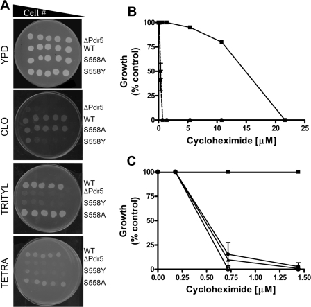
FIGURE 1.
Drug sensitivity of S558Y. A, 2-fold serial dilutions of Pdr5 strains were plated on YPD media or on YPD media containing 15 μm clotrimazole, tritylimidazole, or tetrabutyltin. The number of cells plated ranged from 1.0 × 10⁶ to 0.5 × 10⁴; scans of plates that were incubated for 96 h at 30 °C are shown. A representative figure from three independent experiments is shown. B, quantitative analysis of drug sensitivity was carried out by inoculating 500 cells bearing the WT and mutant Pdr5s in the presence of increasing concentrations of cyh (0.05–21.6 μm). The cells were incubated at 30 °C for 48 h and then quantified by measuring the absorbance at 600 nm. The strains used were R-1 (Δpdr5 = •), R-1 + pSS607 (WT = ▪), and R-1 + pS558Y (▴). Growth at each concentration of cycloheximide is shown as (% control), i.e. growth in the absence of cyh. C, same protocol was used to compare single- and double-copy mutant strains: single-copy WT (JG2000 = ▪), single-copy S558Y (R-1 + pS558Y = ▾), double-copy S558Y (JG2010 = ▴), and R-1 (○). The figures (B and C) show the mean values of three independent experiments, and the error bars represent the S.D.

