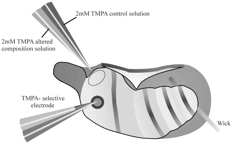
Figure 1.
Schematic diagram showing experimental protocol utilizing ventral approach to the right ear of a guinea pig. TMPA was irrigated across the RWM by a double barreled pipette, allowing remote switching of solution perfused, with excess fluid removed by a wick. The rate of entry of TMPA into scala tympani was measured by sealing a calibrated double-barreled TMPA-selective microelectrode into the basal turn of scala tympani. Cyanoacrylate and silicon glues were used to seal the electrode in place.

