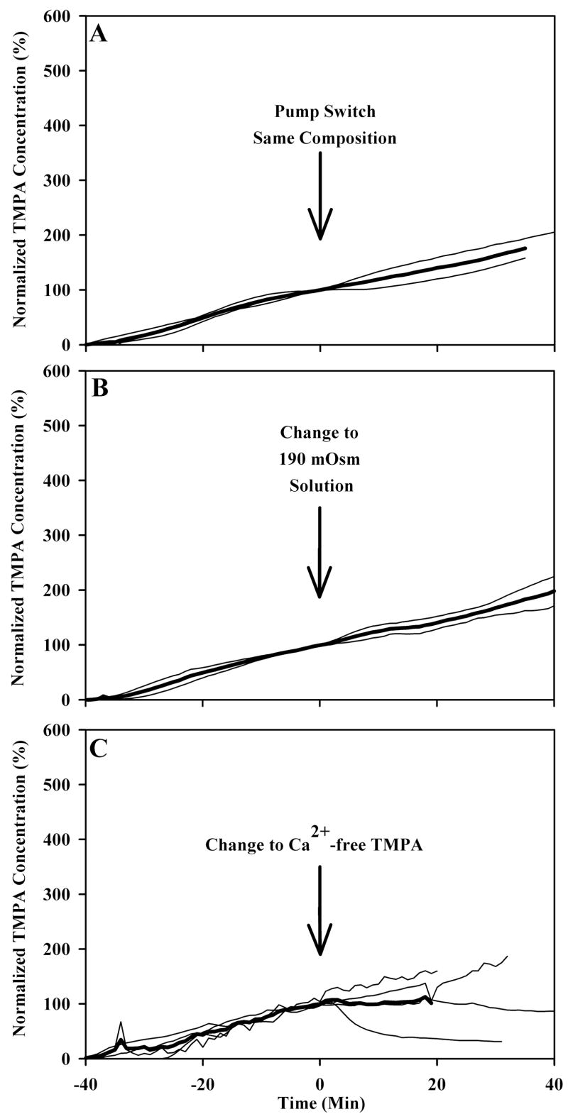
Figure 4.
Several manipulations resulted in no alteration of TMPA entry across the round window. Light traces in panel A, B, and C show individual experiments in which TMPA concentration was measured in the basal turn of scala tympani. For 40 minutes prior to time zero, TMPA in artificial perilymph was irrigated across the round window membrane. At time zero, in panel A, the identical solution was perfused through the second barrel in the double barreled pipette to serve as a control. In panels B and C, at time zero the solution irrigated across the round window membrane was changed to a different composition, as indicated, but containing an identical TMPA concentration. Measured TMPA concentrations were normalized with respect to the concentration in scala tympani prior to the solution change. The mean curve of the group is shown as a heavy line.

