
Fig. 6.


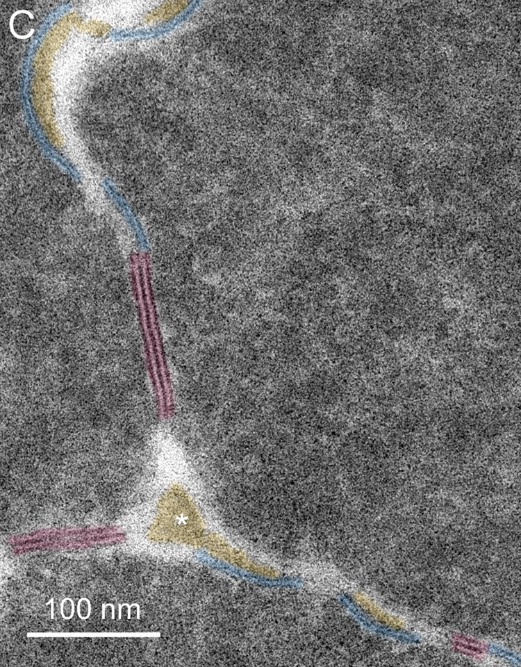
Membranes at cell-cell and cell-process intersections. A. Overview of three cells shows a typical pattern of cellular architecture in a mature nuclear cataract. Numerous edge processes (EP) are displayed at the interfaces of adjacent cells (51 years old). B. High magnification of one region (boxed in A) reveals damaged membranes from three cells and two edge processes. Extracellular space deposits (yellow) appear on curved membranes (blue and green lines) and similarly staining material occurs at trigonal intersections (*). The trigonal points are extended extracellular channels that are partially filled with protein-like material. C. A trigonal intersection in which the protein-like material is triangular in shape (*) and the adjacent membranes and junctions are distinct (70 years old).