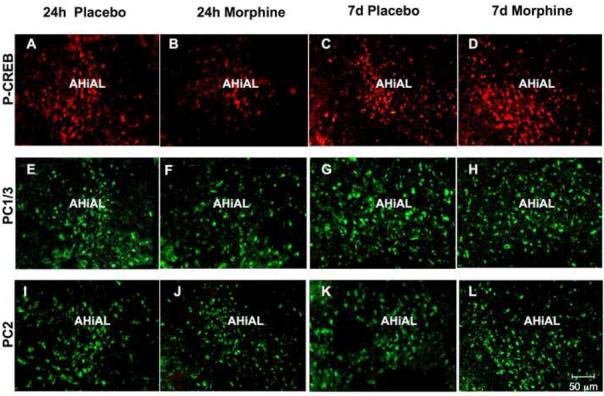
Fig. 5.


Changes in P-CREB, PC1/3 and PC2 levels in the anterolateral nucleus of the amygdala (AHiAL) of rats after short-term or long-term morphine treatment. Placebo or morphine pellets were implanted subcutaneously in the neck of rats for 24 h or seven days. Rats were then sacrificed, perfused with paraformaldehyde and their brains excised and fixed. Coronal sections of frozen slices (8-μM thick) were mounted and immunofluorescence for P-CREB (A-D), PC1/3 (E-H) and PC2 (I-L) was performed. Figures are representative of 4 rats/group analyzed. Quantification of P-CREB (M), PC1/3 (N) and PC2 (O) immunoreactivity is expressed as the mean +/- SEM Integrated Optical Density/area. n=4 rats per group. Magnification 200X. Bar = 50 μM. **p<0.005. P, placebo; M, morphine.
