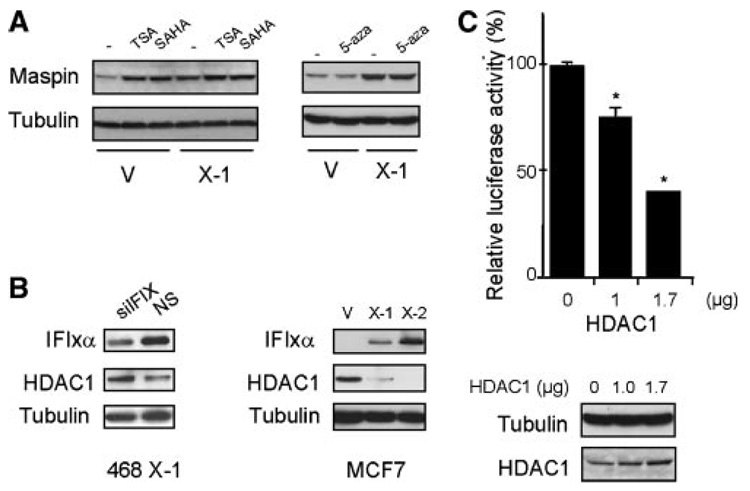
Figure 3.
HDAC1 is negatively regulated by IFIXα. (A) MDA-MB-468 control (V) and IFIXα stable cells (X-1) were treated with 100 nM of TSA, 1 µM of suberoylanilide hydroxamic acid (SAHA), or 250 nM of 5-aza-2′-deoxycytidine (5-aza) for 14 h, and maspin and tubulin expression in both treated and untreated cells was determined by immunoblot analysis. (B) MDA-MB-468 IFIXα stable cells (X-1) were transiently transfected with siRNA against IFIXα (left panel). MCF7 cells were stably expressed with vector control (V) and IFIXα (X-1 and X-2). IFIXα, HDAC1, and tubulin expression were determined by immunoblot analysis. (C) H1299 cells were transiently co-transfected with 0.3 µg of Firefly luciferase plasmids containing maspin promoter, pRL-TK renilla luciferase plasmids, and the indicated amounts of HDAC1 expression plasmids. Twenty-four hours after transfection, the cells were harvested and subjected to a dual luciferase assay (Promega, Madison, WI). Data are expressed as averages ± standard deviation (n = 3). *Significantly different from the control, P < 0.001. The HDAC1 protein expression in H1299 cells are shown below the graph.

