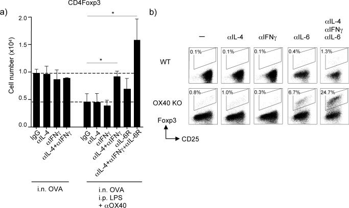
Figure 6. OX40 and the cytokine microenvironment controls the generation of antigen-specific Foxp3+ T cells.
(a) Antigen-specific Foxp3+ cells derived in vivo from adoptively transferred naive wt OT-II CD4 cells were tracked in pooled lymph nodes as described in Fig. 2 under the experimental setting in Fig. 4. Mice were left untreated, or injected with LPS and anti-OX40 i.p. at the time of exposure to soluble OVA i.n., as in Fig 4. Mice were also injected with control IgG, or anti-IL-4, anti-IFN-γ, and anti-IL-6R, alone or together, during the period of tolerance induction. Results are the mean number of Foxp3+ OT-II T cells ± SEM in lymph nodes at day 5, from 4 mice per group. (b) Naive CD25−(Foxp3−) CD4+ cells from WT or OX40 KO AND TCR transgenic mice were cultured in vitro with MCC peptide presented on fibroblast APC that express CD80 and OX40L, in the presence of exogenous TGFβ. Blocking antibodies to IL-4, IFN-γ and IL-6 were added to culture on day 0. After 3.5 days, the primed T cells were stained for the expression of CD25 and Foxp3 after gating on CD4. Data show percent Foxp3+ cells. Results are representative of two experiments.

