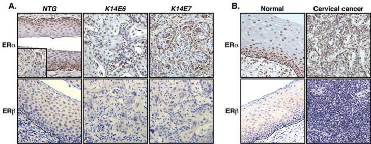
Fig. 1. ERα but not ERβ is detected in cervical cancers.
(A) ERα is expressed in murine cervical cancers. Archival PFA-fixed and paraffin-embedded female reproductive tracts of indicated mice treated with 17β-estradiol were stained for ERα (upper panel) or ERβ (lower panel). Stromal cells stained for ERα is shown in the inset. Shown are representatives of more than six cancers in each transgenic mouse. More than 700 cancer cells in at least three different areas of each cancer were examined for positive staining. Nuclei were counterstained with hematoxylin. Black lines indicate basement membrane separating epithelium from underlying stroma. (B) ERα is expressed in human cervical cancers. Formalin-fixed and paraffin-embedded human cervical tissue sections were stained as described in (A). Shown is representative of three HPV-positive and four HPV-negative cervical cancers.

