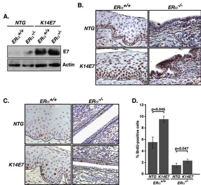
Fig. 4. Functional E7 is expressed in K14E7/ERα−/− cervical epithelium.
(A) E7 is expressed in the cervix of ERα−/− mice. Vaginal tissue lysates from mice of indicated genotype was immunoblotted with anti-HPV-16 E7 or anti-actin antibodies. (B) Mcm7 expression is enhanced in K14E7/ERα−/− cervix compared to NTG/ERα−/− cervix. Paraffin sections of female reproductive tracts from estrogen-treated mice of the indicated genotypes were stained for Mcm7 and nuclei were counterstained with hematoxylin. Shown are representatives of three mice in each genotype. Black lines indicate basement membrane separating epithelium from underlying stroma. (C) BrdU incorporation into DNA is increased in K14E7/ERα−/− cervix compared to NTG/ERα−/− cervix. Paraffin sections of female reproductive tracts from estrogen-treated mice of indicated genotypes were stained for BrdU. Nuclei were counterstained with hematoxylin. (D) Results shown in panel C were quantified (ERα+/+ background, n = 3 for each genotype; ERα−/− background, n = 10 for each genotype). More than 500 cells in five different areas of cervix per mouse were examined. P-values for two-sided Wilcoxon's rank sum test are shown.

