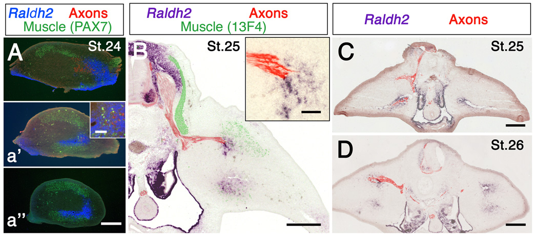
Figure 4.
Relationship between axons and Raldh2 expression in hindlimb. (A-a’’) Raldh2, muscle masses and axons in cross-sections through progressively more distal regions of hindlimb. At St. 24 axons from the sciatic plexus have begun to grow into posterior limb, but have not yet left the more anterior crural plexus. Note that axons are associated with Raldh2 in A and a’, and that Raldh2 extends distal to the axons (a’’). Inset in a’ shows axons and Raldh2 at higher magnification. (B) Raldh2, muscles and axons in a section though spinal cord and limb at the level of dorsal root gangion LS2. Inset shows Raldh2 and axons at higher magnification. (C, D) Raldh2 and axons in embryos in which the right half of the neural tube was removed at St. 17 and St. 16, respectively. Note that axons are missing from denervated limbs, but Raldh2 expression is unchanged. Scale bars: 300 µm except insets. Insets: 50 µm.

