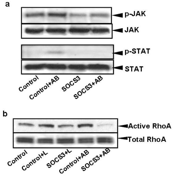
Figure 5. Phagocytosis of apoptotic bodies by HSC induces JAK1 and STAT3 phosphorylation and RhoA GTP-ase activity.
A) LX-2 cells were cultured in serum-free medium, mock-transfected (only pCMV-XL6) or transfected with the SOCS3 construct, and 48 hours later exposed to AB for half an hour. Western blot analyses were performed to detect JAK1 and STAT3 phosphorylation. Transfection of LX-2 cells with the SOCS3 construct inhibited JAK1 and consequently STAT3 phosphorylation. B) RhoA pull-down assay was performed in SOCS3 or mock-transfected LX-2 cells after exposure to either leptin or AB. RhoA activity decreased in SOCS3-transfected cells compared to control (mock transfected cells) after leptin, and especially after AB treatment.

