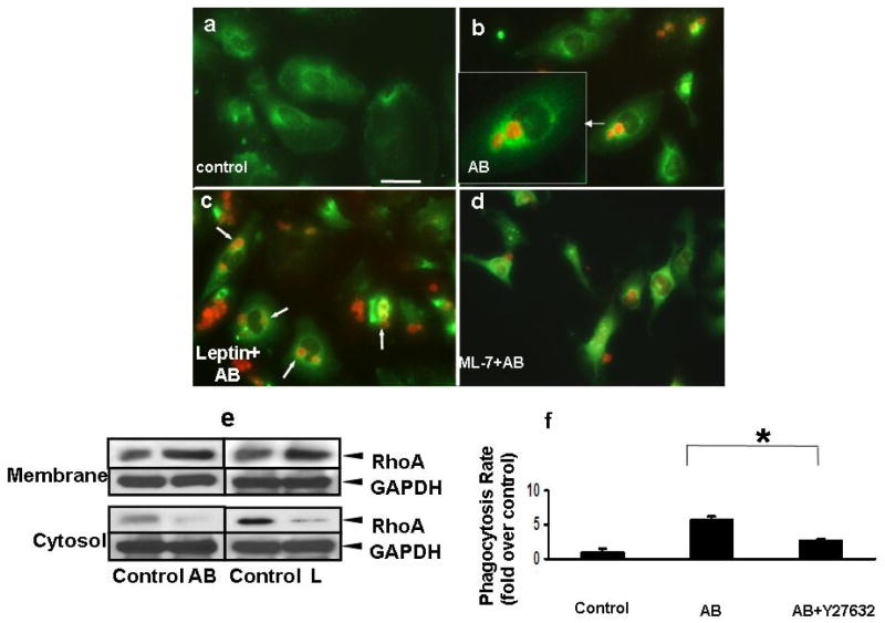
Figure 7. Leptin induces translocation of RhoA to phagosomes and ROCK activation.
LX-2 cells were exposed to AB, leptin plus AB in the presence or absence of ML-7 (AB were labeled previously with TAMRA, red). a) Immunofluorescence was performed and RhoA (green) was seen in the cytoplasm of control cells, and b) translocating to the phagosomes (insert) in AB, or c) L+AB treated cells (arrowheads). In the presence of leptin, more RhoA positive phagosomes were seen. d) After treatment of a MLCK inhibitor ML-7, less phagocytosis was observed and RhoA failed to localize to the phagosomal membrane around AB.
e) Western blot assay was performed to assess membrane-bound RhoA. RhoA protein was increased in the membrane fraction and decreased in the cytosol after the exposure to either AB or leptin. GAPDH served as equal loading control. (N=3)
f) Phagocytosing LX-2 cells were exposed to the ROCK inhibitor Y27632 (10uM) prior to exposing them to AB, and this decreased the phagocytic rate by 0.53-fold when compared to that without the inhibitor. Mean ±SE, N=3, * p<0.05

