
Figure 1.
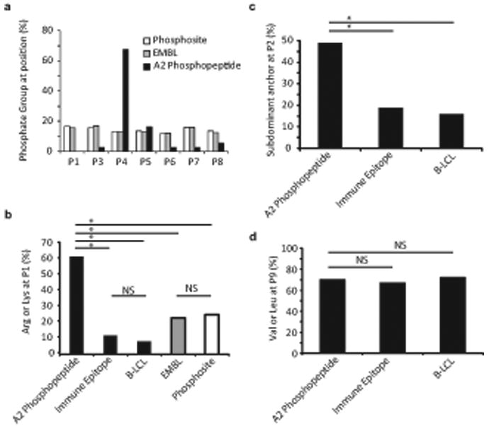
Bioinformatic characterization of the HLA-A2–restricted phosphopeptide repertoire. (a) Distribution of phosphorylated residues among naturally processed (A2 phosphopeptide) and predicted HLA-A2 binding phosphopeptides (Phosphosite, EMBL). The frequency of phosphorylated residues at each position is displayed for naturally processed HLA-A2 associated phosphopeptides, and for peptides in EMBL and Phosphosite datasets that contain phosphorylation sites and are predicted, according to criteria described in Methods, to bind HLA-A2. (b) Representation of positively charged residues (Arg or Lys) at P1 among naturally processed HLA-A2 associated phosphopeptides, phosphopeptides from the EMBL or Phosphosite datasets that are predicted to bind HLA-A2 and contain a p-Ser residue at the P4 position, and datasets of naturally processed non-phosphorylated peptides (B-LCL) and known HLA-A2 binding peptides (Immune Epitope). Selection criteria for the latter two datasets are described in Methods. * = P<0.001, NS= not significant. (c, d) Representation of subdominant residues at the P2 anchor position (c) and the PC (P9) position (d) in naturally processed HLA-A2 associated phosphopeptides and in datasets of naturally processed non-phosphorylated peptides and known HLA-A2 binding peptides.