
Figure 3.
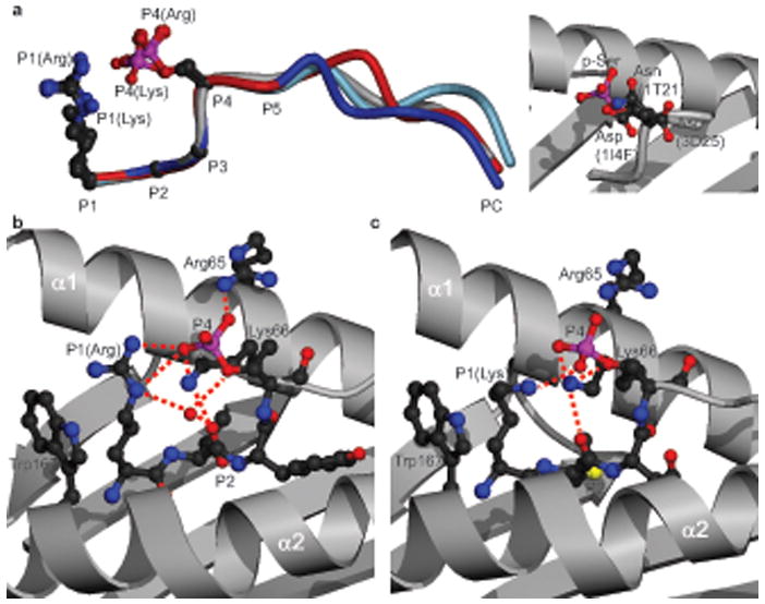
Interactions of the P4 p-Ser with the HLA-A2 heavy chain. (a) Left, superposition of the 4 phosphopeptide structures (RTY (blue), KMD (red), PKD2 (grey), RQA_M (light blue)) on the basis of P1 to P4 Cα atoms, with P1 and P4 sidechains shown. Right, orientation of the PKD2 P4 p-Ser side chain (red and pink) relative to P4 sidechains (Asp, Asp, and Asn) of non-phosphorylated HLA-A2-restricted peptides: (PDB codes 1I4F, 3D25, and 1T21 respectively). (b, c) Interactions of the RTY (b) and KMD (c) p-Ser moieties with HLA-A2 α1-α2 helices. Red dashed lines, hydrogen bonds; red sphere, conserved water molecule.