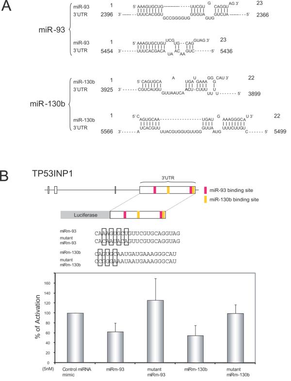
Figure 3. miR-93 and miR-130b target the 3′UTR of TP53INP1.

A) Schematic representation of miR-93 and miR-130b targets in the 3′UTR of TP53INP1. The positions of the miRNA binding sites correspond to the location of the GenBank sequence NM_033285. B) The 3′UTR of TP53INP1 was PCR-amplified and then cloned downstream a firefly luciferase gene (upper panel). The resulting construct was used to verify the inhibitory activity of miR-93 and miR-130b by transfecting miRNA mimics (miRm-93 and miRm-130b) (lower panel). Specificity of the inhibition was determined by transfecting miRNA mimics with or without the indicated mutations (boxed) in the seed sequences (middle panel). A CMV-driven renilla luciferase construct was co-transfected as a normalization control for firefly luciferase activity.
