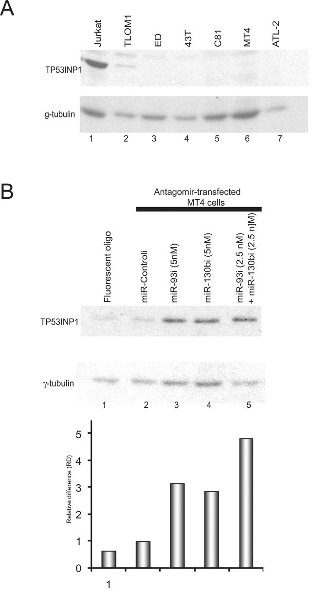
Figure 4. Induced expression of TP53INP1 by transfection of antagomirs.
A) Western blot analysis detected endogenous TP53INP1 expression in Jurkat cells, but not in HTLV-1 transformed cell lines (TLOM1, ED, 43T, C81, MT4 and ATL-2) (Upper panel). Immunoblotting of γ-tubulin was performed as loading controls (Lower panel). B) Tranfection of antagomirs into MT4 cells targeting the cell endogenous miR-93 and miR-130b increased TP53INP1 expression. Immunoblotting of γ-tubulin was performed as a loading control. miR-93 and miR-130b antagomirs are labeled as miR-93i and miR-130bi. Bottom panel shows quantification of the relative intensities of the TP53INP1 bands after normalization to the corresponding tubulin signals. Proliferation and apoptotic assays shown in Figure 5A and B were done in parallel with this experiment using the same set of cell samples (see Figure 5A and B).

