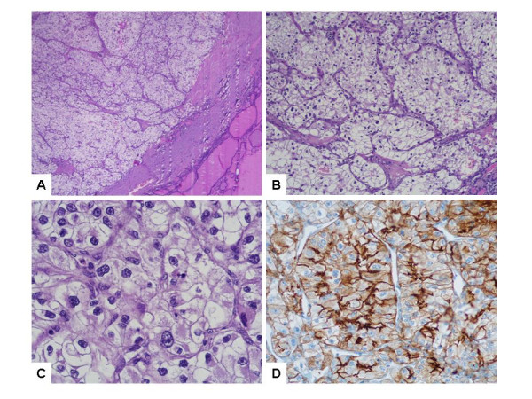
Figure 1.

Histopathology of renal cell carcinoma metastasis in the thyroid: capsulated intrathyroidal nodule (A) composed of nests and cords of large clear cells (B) with abundant optically empty cytoplasm, sharply outlined boundaries and moderately atypical nuclei (C). The clear cells are CD10-immunoreactive (D). (H&E, ×10, ×100 and ×400; avidin-biotin-peroxidase, ×400).
