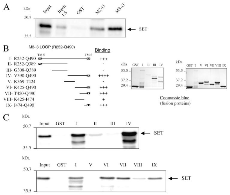
FIGURE 5. Localization of the site of SET interaction with the M3-i3 loop.
A, the purified GST fusion proteins encompassing the i3 loop of the M2- or M3-MRs were incubated with recombinant His-tagged SET, and the bound proteins were eluted, separated on denaturing polyacrylamide gels (10%), and immunoblotted with a polyclonal anti-SET antibody. B, the M3-MR Arg252—Gln490 i3 loop peptide was progressively truncated at the amino and carboxyl termini to generate different M3-i3 loop fragments II–IX. Each construct was expressed in BL21 bacteria, and the purified GST fusion proteins were electrophoresed on denaturing polyacrylamide gels (10%) and visualized by Coomassie Blue staining. C, each of the M3-i3 constructs (B) were evaluated in protein interaction assays with recombinant SET as described in the legend to Fig. 4.

