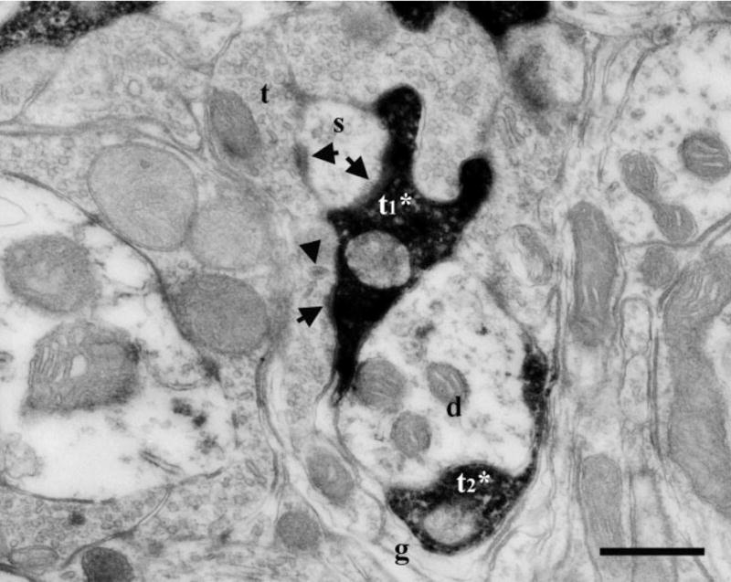
Fig. 2.

Electron micrographs of DAB-labeled terminals in the dorsal zone of the NTS of a sodium-restricted rat. A labeled GSP terminal (t1*) engaged in a synaptic triad and formed asymmetric synapses (arrows) on both a spine (s) and an unlabeled terminal (t). The unlabeled terminal (t) contained a dense-cored vesicle (arrowhead) and formed a synapse (arrow) on the same spine (s) as the labeled terminal (t1*). Glia (g) surrounded the entire arrangement including an additional labeled terminal (t2*) and a dendrite (d). Scale bar = 0.5 μm.
