Fig. 5.

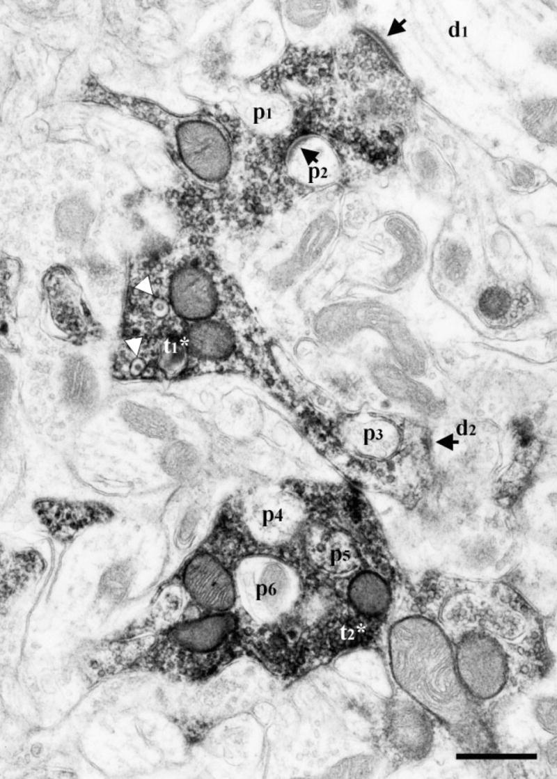
Electron micrograph of DAB-labeled CT terminals in the dorsal zone of the NTS of a sodium-restricted rat. Two labeled terminals (t1*, t2*) encased many protrusions (p1–p6) emerging from surrounding neuropil. The protrusion p6 is identified as a dendrite shaft, based on presence of mitochondria. One of the labeled terminals (t1*) contained dense-cored vesicles (white arrowheads) and formed synapses (arrows) with two separate dendrites (d1, d2) and with an encased protrusion (p2). Scale bar = 0.5 μm.
