
Figure 4.
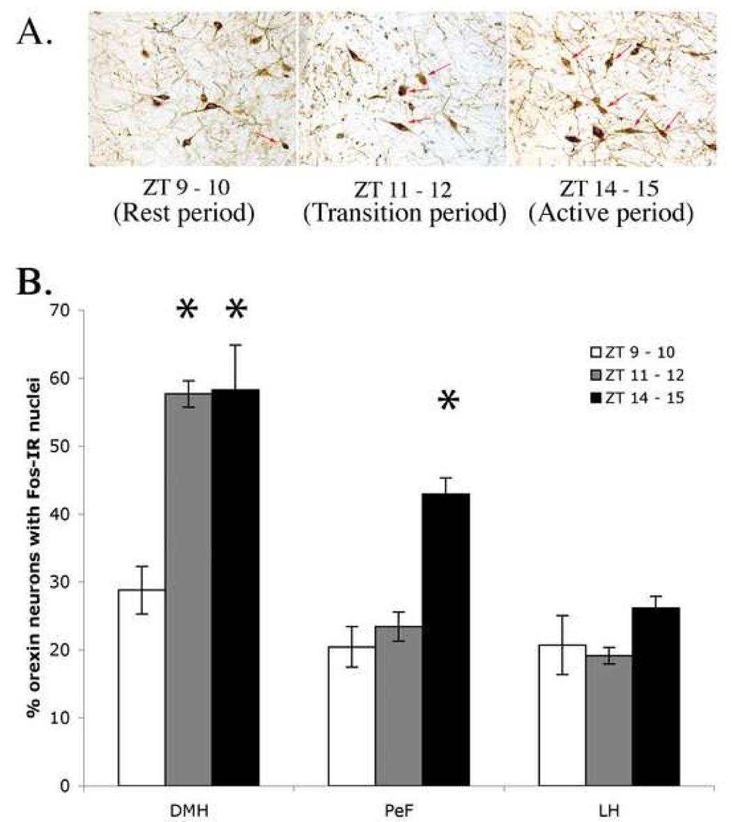
A, FOS expression in orexin-positive neurons in animals sacrificed 90 minutes after ZT 9–10, 11–12, or 14–15 in the dorsomedial hypothalamus. B, average counts of orexin-IR neurons that were also Fos-IR at ZT 9-10 (white bars), 11–12 (grey bars) and 14–15 (black bars) in the dorsomedial hypothalamus (DMH), perifornical (PeF), and lateral hypothalamus (LH). Fos-IR in orexin neurons was elevated during the active period compared to the rest period in the DMH and PeF, but not significantly in the LH. Only orexin neurons located in the DMH showed an increase in Fos immunoreactivity over ZT 9–10 at ZT 11–12 and neither PeF nor LH orexin neurons showed a similar increase. Asterisks denote significant differences from rest.