
Figure 5.
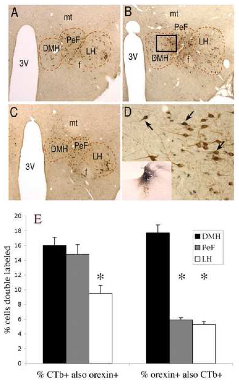
Double labeling of orexin afferents to the LC. A – C, Frontal sections at low-power through different levels of the rat hypothalamus (ordered caudal to rostral) double stained for orexin (brown) and CTb (retrogradely transported from the LC, dark blue). These images depict the different areas that were defined for counting orexin neurons: DMH, PeF and LH. Abbreviations: 3V – third ventricle; mt – mammillothalamic tract; f – fornix. D, High-power photo taken from box in panel B,showing doubly labeled neurons in the DMH and PeF regions (at arrows). Inset: example injection site of CTb in the LC. E: Bar graph summarizing counts of neurons labeled for orexin, CTb, or both. Note that most LC-afferent orexin neurons are located in the DMH and PeF with fewer in the LH. In addition, a igher percentage of orexin neurons in the DMH contain CTb, indicating that they project to LC, than in the PeF or the LH. Asterisks denote significant differenes (p <0.05) from DMH.