Figure 3.
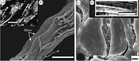
SEM of collagen fibre bundles from the white shark C. carcharias. (a) Degraded and dehydrated collagen fibre bundles showing beaded structure. (b) High resolution of (a): shows fibres within the collagen fibre bundle; the waviness indicates loss of tension (see text). The waves transfer to the fibre bundle and ultimately create the beaded structure. (c) Fresh collagen fibre bundle in cross section. The fibre bundle is fractured down the middle, showing fibres still retaining original tension. (d) Component fibrils from the fibre showing 67 nm axial banding. Scale bars, (a) 500 μm, (b) 50 μm, (c) 10 μm and (d) 1 μm.

