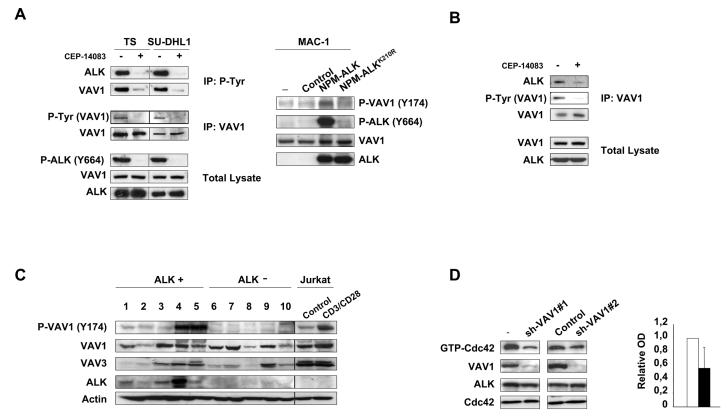
Figure 3. NPM-ALK phosphorylates VAV1 and activates Cdc42 through VAV1 phosphorylation.
A, left panel: TS and SU-DHL1 cells were cultured with 300nM ALK inhibitor CEP-14083 for 6 hours and with orthovanadate for 10 min before harvesting. Total cell lysates were immunoprecipitated with anti-VAV1 or anti-P-Tyr antibodies and blotted with the indicated antibodies. Right panel: MAC-1 cells were transduced with empty vector, NPM-ALK or NPMALKK210R retroviruses. Total cell lysates were blotted with the indicated antibodies. B, SU-DHL1 cells were cultured with 300nM ALK inhibitor CEP-14083 for 6 hours and with orthovanadate for 10 min before harvesting. Total cell lysates were immunoprecipitated with anti-VAV1 antibody and blotted with the indicated antibodies. C, frozen tissues from primary ALK positive (lanes 1-5) and negative (lanes 6-10) ALCL cases were lysed and blotted with the indicated antibodies. Jurkat T cells were stimulated with CD3 and CD28 for 10 min as a control for VAV1 phosphorylation. D, wild-type TS cells were transduced with two different lentiviral contructs encoding for specific sh-RNAs against VAV1 or a control sequence. Total cell lysates were used for Cdc42 pull-down assay or blotted with the indicated antibodies. The results are from one representative experiment. Histograms indicate the quantification by optical densitometry as described above.

