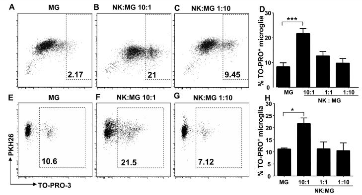
Figure 1. Natural Killer cells kill human microglia.
Fetal human microglial cells were cocultured with allogeneic (upper panel) or autologous (lower panel) IL-2 activated NK cells for 4 hr. The extent of cytotoxicity was quantified by the relative number of live human microglial cells labeled with PKH-26 and dead, permeabilized cells labeled with both PKH-26 and TO-PRO-3 iodide. Fig. 1 A, E: spontaneous permeabilization of primary human microglia. Fig. 1 B, F and C, G: cellular permeabilization at different E:T ratios. Fig. 1 D, H: diagrams summarize at least three experiments and display means and SEM of TO-PRO-3+ microglia cells compared to PKH26+ microglial cells after coculture with increasing E:T ratios. D: MG vs 10:1, *** p<0.0001 for E:T ratio of 10:1. H: MG vs 10:1, * p=0.0492 for E:T ratio of 10:1, student t test. MG: Microglia, NK: Natural Killer cells.

