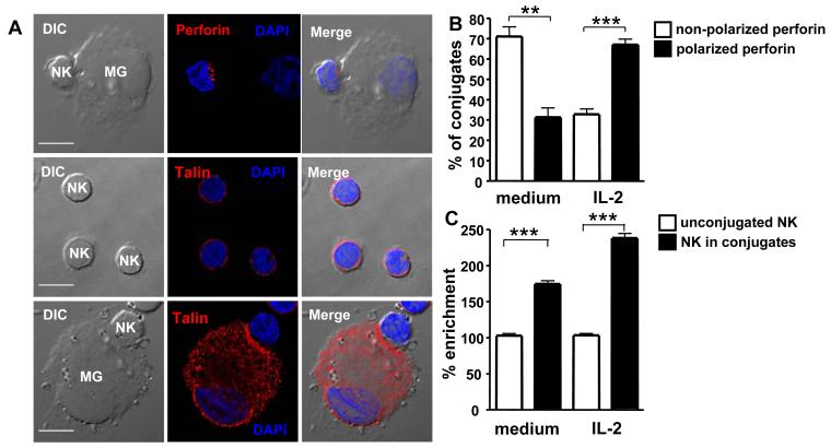
Figure 3. Synapse formation and perforin polarization in NK cell-microglia conjugates.
Polarization of the cytoskeletal protein talin, indicative of synapse formation, and of the cytotoxic molecule perforin in NK cells was determined by 2-dimensional immunofluorescence microscopy after 1 min. Fig. 3A: Perforin and talin polarization in IL-2 activated NK cells conjugated to microglia. Fig. 3B: Perforin is polarized in conjugates of microglia with IL-2 activated NK cells. Non-polarized vs. polarized perforin in conjugates with resting NK cells, ** p=0.001; Non-polarized vs. polarized perforin in conjugates with activated NK cells, *** p<0.0001; student t test. Fig. 3C: relative enrichment (RE) of talin to cell contact areas in NK cell conjugates with microglia compared to NK cells alone. Resting NK cells alone vs. conjugated, *** p<0.0001; Activated NK cells alone vs. conjugated, *** p<0.0001; student t test. Representative and summarized data for three experiments are shown.

