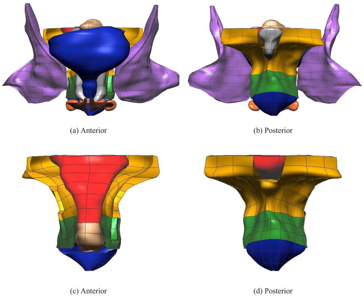
Fig. 1.
The final fitted models from the MRI data set. Shown are (a) anterior and (b) posterior views showing 12 of the 13 components – levator ani (LA) (gold), puborectalis (PR) (green), external anal sphincter (EAS) (blue), internal anal sphincter (IAS) (beige), rectum (red), transverse perineae (orange), perineal body (orange), coccyx (silver), uterus (beige), vagina (silver), obturator internus (purple) and bulbospongiosus (brown). Also shown are enlarged views of the (c) anterior and (d) posterior of the LA, PR, EAS, IAS and rectum. For clarity the lumen has not been shown in these views.

