
Fig. 1.
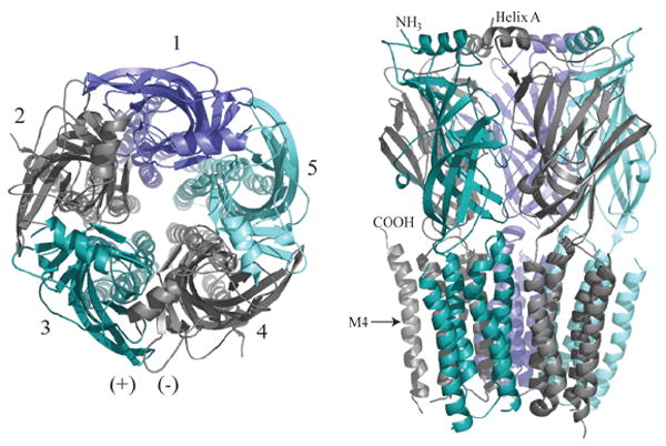
Molecular model of Cys-Loop pentameric receptor. The core GABAAR homology model was constructed as described by Mercado and Czajkowski (51) using Sybyl 7. 1 (Tripos Inc., St. Louis, MO). Left: Top view, as if from the synaptic cleft. Subunits are labeled 1-5 counterclockwise with the principal subunit colored slate (subunits 2 and 4). For the GABAAR, subunits 1-5 correspond to γβαβα, although position 1 can also be occupied by other subunits, such as δ, ε, θ, or π (2); for the muscle nAChR, 1-5 are βαγαδ or βαεαδ; for the neuronal nAChRs and glyRs, 1-5 are (β/α)αβαβ (the first position is still in dispute for glyRs); for 5-HT3Rs 1-5 are BABAB. One (+)/(-) interface is illustrated. Right: A sidelong view shows the extracellular N-termini (mostly β-strands, and one α-helix denoted Helix A) resting atop the transmembrane α-helical spans M1-M4. M4 is labeled to show the position of the C-terminus. The intracellular loop between M3 and M4 is not included, for clarity.