
Fig. 2.
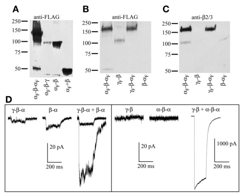
Western blots of several GABAAR concatamers. All constructs shown include signal peptide in the linker region. (A) Blot probed with anti-FLAG M2 monoclonal antibodies (Sigma). Subunits and tandems were immunoprecipitated from HEK293 cells transfected with α FLAG-tagged α1-β2-α1 (Lane 1), α1-β2-γ2 (Lane 2), α1-β2 (Lane 3), or free α1 plus β2 (Lane 4). The triple concatamer is expected at approx 165 kD, the tandems at approx 110 kD, and the monomers near 50–55 kD. The first lane was overloaded to show breakdown products, presumably free FLAG-tagged α1-subunit. Lane 2 shows no signal at 165 kD for αF-β-γF but, rather, a fragment that runs at the size of a double subunit tandem. Conversely, Lane 3 shows a robust signal for αF-β. (B) Blot probed with anti-FLAG antibody again showing breakdown of αF-β-αF (Lane 1), but full-length γF-β (Lane 2) and γF-β-αF (Lane 3) with no apparent breakdown, and no signal for β-αF (Lane 4). Note that both anti-FLAG and anti-β2 antibodies are less efficient out of first position. (C) Same blot as in (B) reprobed with polyclonal antibodies that recognize β2 or β3. Lane 1 shows a product of 110 kD, presumably β-αF as a second breakdown product of αF-β-αF. Lane 4 shows low expression of β-αF. The same result was obtained multiple times with multiple β-αF constructs, whereas γF-β constructs expressed better in other experiments, with no appreciable breakdown (data not shown). (D) Representative traces from whole-cell recordings of HEK293 cells transfected with tandem constructs, alone or in combinations designed to yield γβαβα pentamers. All constructs include 9Q (for the α-β linker) or 10Q (all others) plus signal peptide. Each trace shows current from exposure to 10 mM of GABA for 20 ms, and current scale bars for all recordings are 20 pA, with the exception of “γ-β + α-β-α,” which is 1000 pA. Left: γ-β-α and β-α tandems exhibit currents when expressed alone. Two traces for “γ-β-α + β-α” are overlaid to show range, and the rather small amplitude of the currents. Right: In at least eight cells each, neither γ-β nor α-β-α showed any current when expressed on their own, whereas robust currents were seen when these two constructs were co-expressed at a 1:1 ratio. (Portions of these data are reprinted with permission (11); Copyright 2005 by the Society for Neuroscience.)