Figure 1.
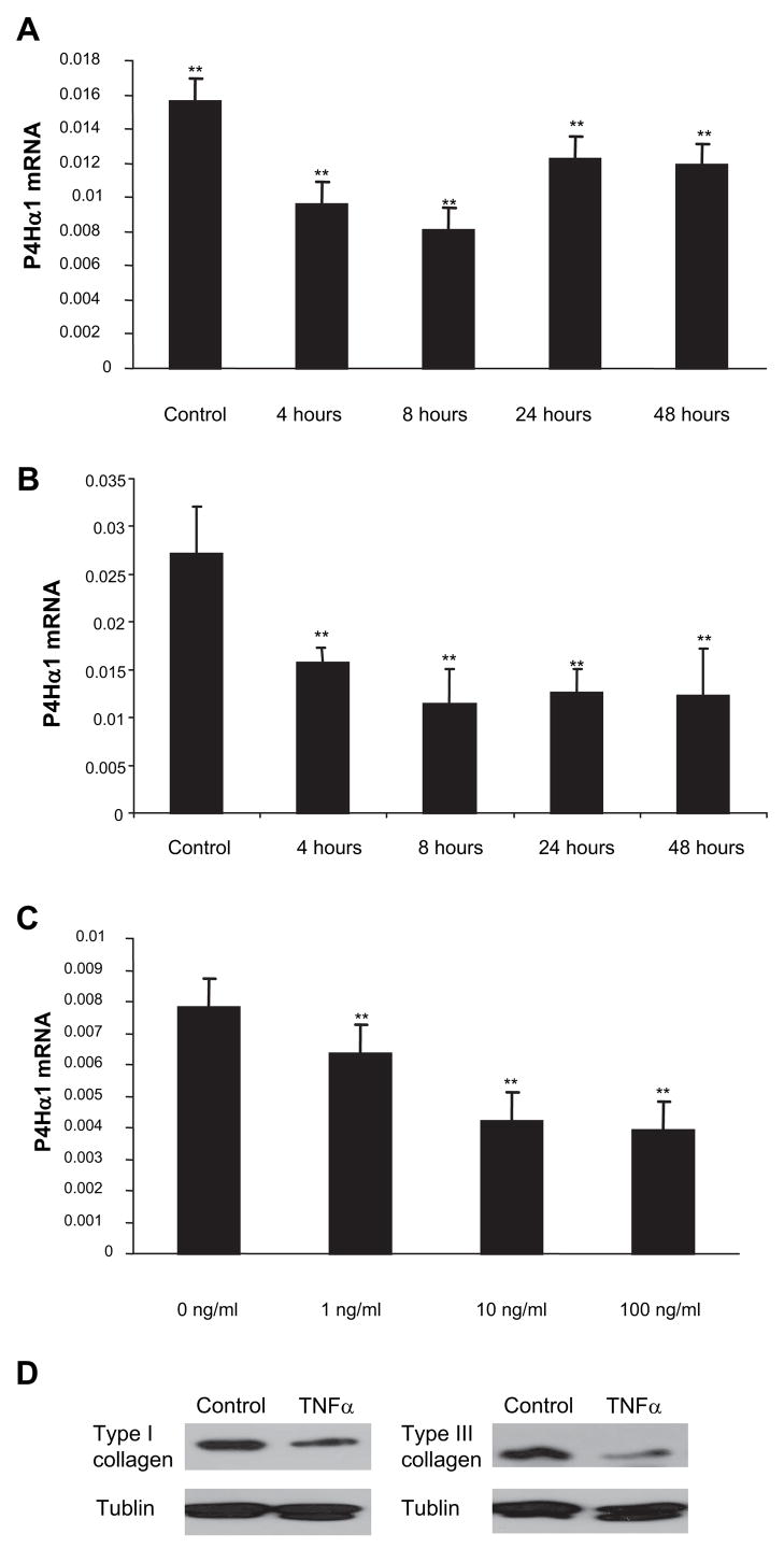
TNF-α suppresses P4Hα1 mRNA expression in HASMCs. Quantitative realtime RT-PCR was used to measure the mRNA levels. A, Levels of P4Hα1 mRNA were measured from HASMCs treated with 100 ng/mL TNF-α for 0, 4, 8, 24, and 48 hours. B, HASMCs were treated with 100 ng/mL TNF-α at 0 hours and replenished with fresh 100 ng/mL TNF-α at every 8 hours till 48 hours. Levels of P4Hα1 mRNA were measured at 0, 4, 8, 24, and 48 hours. C, HASMCs were treated with 0, 1, 10, and 100 ng/mL TNF-α for 8 hours. ANOVA was used to compare the among-group differences in both the time course and dose-dependent effects in TNF-α-mediated P4Hα1 inhibition. **P<0.01 indicates the comparison between the treated cells and the control cells in the Bonferroni post hoc between group analyses. D, Western blot of type I and III collagen from HASMCs treated with 100 ng/mL TNF-α for 8 hours. The total cellular proteins were extracted and measured using Bradford method. Equal amount of proteins were then loaded to 8% SDS-PAGE gel for electrophoresis. ECL chemiluminescent method was used for the type I and III collagen detection using anti-human type I and III antibodies.

