
Figure 1. Expression of OATP1A2 and PXR comparing non-neoplastic and neoplastic breast tissue.
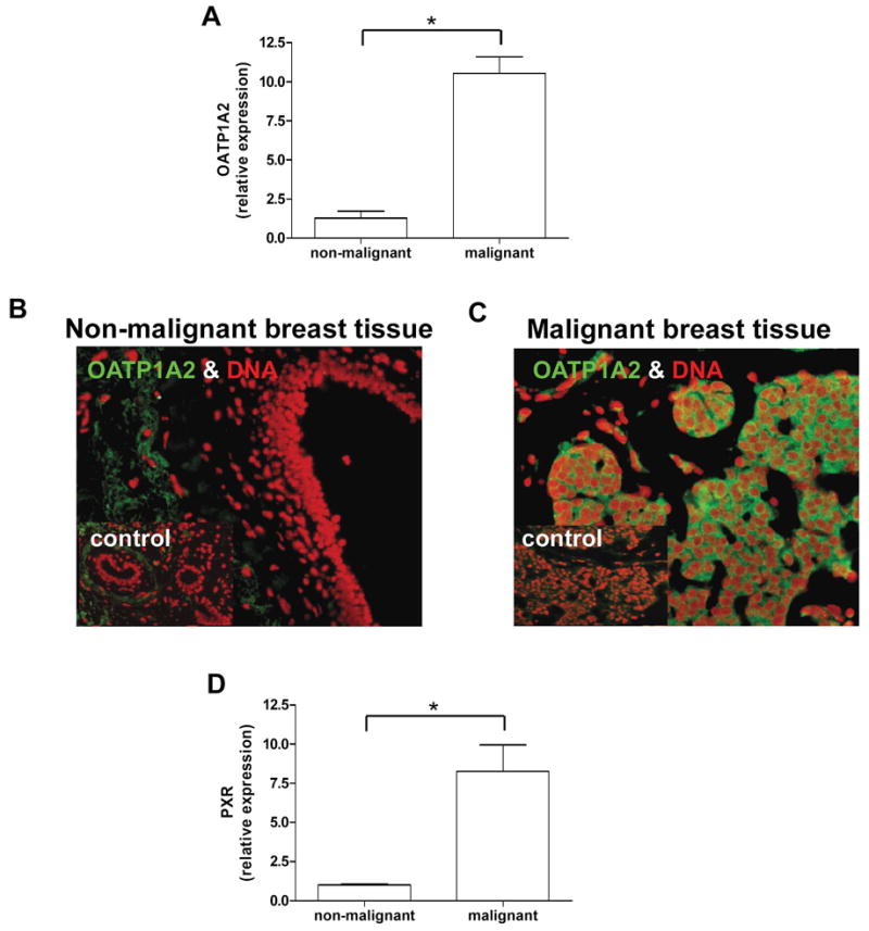
OATP1A2 (1A) and PXR (1C) mRNA-expression was assessed in neoplastic and adjacent non-neoplastic breast tissue samples of 4 individuals using real-time PCR revealing significant higher expression of both genes in the cancerous tissue. Immunofluorescent staining of the uptake transporter OATP1A2 (green) in human malignant and non-malignant breast tissue showed an intense and restricted expression in malignant cells (right panel 1B). No distinct staining pattern was detected in non-malignant breast tissue (left panel 1B). DNA-chromatin was counterstained with DAPI (red). As control the primary antibody was omitted (insert 1B). Data are expressed as mean ± SD, * p<0.05, t-test.