Figure 1.

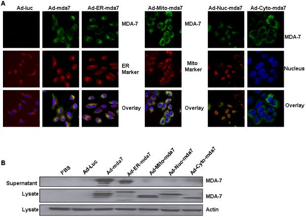
Immunofluorescent confocal microscopic analysis of intracellularly targeted mda-7 adenoviral vectors. A, Analysis of antibody against MDA-7 stained green, endoplasmic reticulum (ER) stained red with ER-Tracker™ dyes, and nuclei stained blue with DAPI demonstrated colocalization of both MDA-7 protein and the ER marker in Ad-ER-mda7-treated A549 cancer cells 72 hours after transfection. Analysis of antibody against MDA-7 stained green, mitochondria (Mito) stained red with Mito Tracker Deep Red 633 (M-22426), and nuclei stained blue with DAPI demonstrated colocalization of both MDA-7 protein and the Mito marker in Ad-Mito-mda7-treated A549 cancer cells 72 hours after transfection. Analysis of antibodies against MDA-7 stained green and nuclei (Nuc) stained red or blue with DAPI demonstrated nuclear localization of MDA-7 protein in Ad-Nuc-mda7-treated A549 cancer cells 72 hours after transfection. Analysis of antibodies against MDA-7 stained green and nuclei (Nuc) stained blue with DAPI demonstrated cytosolic localization of MDA-7 protein in Ad-cyto-mda7-treated A549 cancer cells 72 hours after transfection. B, Western blot analysis of MDA-7 expression in A549 cell lysates and supernatant 72 hours after treatment with PBS, Ad-Luc (2500 vp/ or 24.5 moi), Ad-mda7 (2500 vp/ or 25.5 moi), Ad-ER-mda7 (2500 vp/ or 24.27 moi), Ad-Mito-mda7 (2500 vp/ or 22.1 moi), Ad-Nuc-mda7 (2500 vp/ or 26.59 moi), or Ad-Cyto-mda7 (2500 vp/ or 24 moi). β-Actin expression was analyzed as a control.
