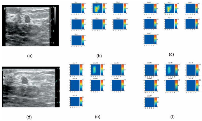
Fig. 10.
(a) Coregistered ultrasound image, (b) the corresponding absorption map obtained at 780 nm, and (c) total hemoglobin map obtained from 53-year-old woman using the black angled probe shown in Fig. 1(a). (d) Coregistered ultrasound image, (e) the corresponding absorption map obtained at 780 nm, and (f) total hemoglobin map using the same probe with the aluminum cover. In absorption and total hemoglobin maps, each slice corresponds to a spatial image of 9 cm×9 cm obtained at 0.3 cm underneath the skin surface to 3.3 cm deep toward the chest wall with 0.5-cm spacing in depth. The fitted background absorption and reduced scattering coefficients from the normal side of the breast were 0.01, 3.78 cm−1 (780 nm), and 0.012, 4.23 cm−1 (830 nm), respectively. The core biopsy result revealed a benign fibroadenoma.

