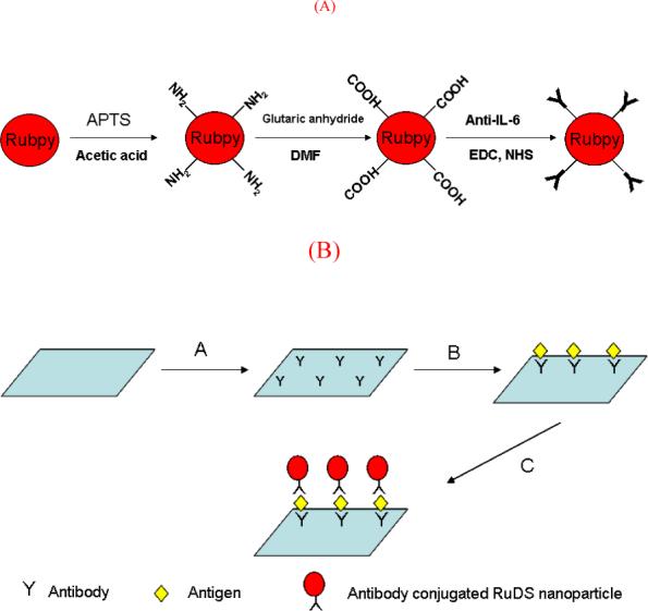
Fig. 3.

(a) Schematic illustration of the procedure for the preparation of antibody conjugated RuDS nanoparticles. (b) The scheme of the RuDS label-based fluorescent immunoassay of IL-6 on a protein microarray format. A) capture anti-IL-6 antibody was printed on the slide. B) antigen, IL-6, was attached to the slide via antibody/ antigen recognition. C) anti-IL-6 antibody-RuDS conjugates was coated on the slide to form a sandwich immunocomplexes with RuDS as tags.
